Factors Associated with Lower Extremity Dysmorphia Caused by Lower Extremity Lymphoedema
T. Yamamoto, N. Yamamoto, H. Yoshimatsu, M. Narushima, I. Koshima. Eur J Vasc Endovasc Surg (2017) 54, 69e77.
Click to read the abstract
Factors Associated with Lower Extremity Dysmorphia Caused by Lower Extremity Lymphoedema.
- Yamamoto, N. Yamamoto, H. Yoshimatsu, M. Narushima, I. Koshima. Eur J Vasc Endovasc Surg (2017) 54, 69e77.
Objectives: Indocyanine green (ICG) lymphography has been reported to be useful for the early diagnosis of lymphoedema. However, no study has reported the usefulness of ICG lymphography for evaluation of lymphoedema with lower extremity dysmorphia (LED). This study aimed to elucidate independent factors associated with LED in secondary lower extremity lymphoedema (LEL) patients.
Methods: This was a retrospective observational study of 268 legs of 134 secondary LEL patients. The medical charts were reviewed to obtain data of clinical demographics and ICG lymphography based severity stage (leg dermal backflow [LDB] stage). LED was defined as a leg with a LEL index of 250 or higher. Logistic regression analysis was used to identify independent factors associated with LED.
Results: LED was observed in 106 legs (39.6%). Multivariate analysis revealed that independent factors associated with LED were higher LDB stages compared with LDB stage 0 (LDB stage III; OR 17.586; 95% CI 2.055e 150.482; p¼ .009) (LDB stage IV; OR 76.794; 95% CI 8.132e725.199; p< .001) (LDB stage V; OR 47.423; 95% CI 3.704e607.192; p¼ .003). On the other hand, inverse associations were observed in higher age (65 years or older; OR 0.409; 95% CI 0.190e0.881; p¼ .022) and higher body mass index (25 kg/m2 or higher; OR 0.408; 95% CI 0.176e0.946; p¼ .037). Conclusions: Independent factors associated with LED were elucidated. ICG lymphography based severity stage showed the strongest association with LED, and was useful for evaluation of progressed LEL with LED.
Main findings
- This fascinating article provides the reader with insight into assessing lymphatic function via ICG lymphography across the different stages of secondary lower limb lymphoedema.
- It included two hundred and sixty eight limbs of 134 female patients with lower extremity lymphoedema secondary to pelvic cancer treatments who underwent bilateral pedal ICG lymphography.
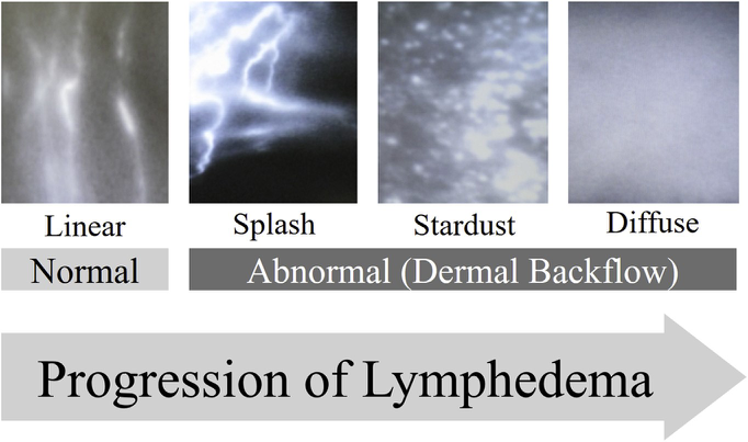
- Leg dermal backflow (LDB) increased as the stage of lymphoedema progressed. Leg dermal backflow was associated with splash, stardust and diffuse presentations on lymphography as seen in the figure below.

Figure 1. Indocyanine green (ICG) lymphography findings. With progression of lymphoedema, ICG lymphography finding changes from Linear pattern (left), to Splash pattern (centre left), to Stardust pattern (centre right), and finally to Diffuse pattern (right).
- The authors found the following ICG lymphography findings for each LDB stage:
- Stage 0 – linear pattern only
- Stage I – linear pattern + splash pattern (around the groin)
- Stage II – linear + stardust pattern (1 region)
- Stage III – Linear pattern + stardust pattern (2 regions)
- Stage IV – Linear pattern + stardust pattern (3 regions)
- Stage V – Stardust pattern (associated with Diffuse pattern)
Lower extremity was divided into three regions: the thigh, the lower leg, and the foot.
- Subclinical lymphoedema on ICG lymphography is seen as a splash pattern and the lymph backflow is reversible.
- When lymphatic collateral pathways fail to compensate for lymph overload, irreversible lymph backflow occurs, and leads to symptomatic progressive lymphoedema; this irreversible lymph backflow is seen as a stardust or diffuse pattern on ICG lymphography.
- The paper indicates this is the first report that has clarified independent factors associated with lower extremity dysmorphia (LED). Indocyanine green (ICG) lymphography based leg dermal backflow (LDB) stage has the strongest association with LED, and is useful for evaluation of late stage lymphoedema with LED. Patients with higher LDB stage should be carefully followed with consideration of aggressive treatment to prevent lymphoedema progression.

