
Breast Oedema After Breast Cancer Treatment
What research from 2019–2026 tells us
About this review: This article is a narrative synthesis of key peer-reviewed research published from 2019 to 2026, with a particular focus on breast oedema following breast-conserving surgery and radiotherapy. It is intended to highlight developments relevant to clinical recognition, assessment and management rather than provide a systematic review of all published studies.
Terminology: In this article, breast oedema is used as the broader clinical descriptor for post-treatment swelling and tissue-fluid change. Some studies specifically use the term breast lymphoedema, particularly where persistent oedema or lymphatic dysfunction is being investigated. Acute postoperative or radiotherapy-related oedema and persistent breast lymphoedema may overlap but are not necessarily synonymous.
“Breast edema is characterized by skin changes, hardness of the breast and pain, but can also be present without visible swelling.” [24]
This observation from Verbelen et al. highlights one of the important advances in our understanding of breast oedema: it is increasingly recognised as more than visible swelling.
Research published from 2019 to 2026 has built a much clearer picture of how breast oedema may present, how it changes over time, factors associated with its development and its impact on women. Assessment has also advanced through breast-specific patient-reported measures, ultrasound, tissue dielectric constant (TDC) and lymphatic imaging, while a growing number of intervention studies are beginning to inform management.
Much of the research reviewed here relates specifically to breast oedema following breast-conserving surgery and radiotherapy. Important questions remain, particularly around consistent diagnostic criteria and optimal treatment, but clinicians now have considerably more information to support recognition, assessment and management as a result of the research published from 2019–2026.[23–26]
More than swelling
One of the clearest messages from recent research is that breast oedema has a broader presentation than breast enlargement alone.
Studies describe heaviness, pain or tenderness, skin thickening, peau d’orange, pitting, redness, enlarged pores and hardness or fibrosis, and breast oedema may be present even when visible swelling is not obvious.[23–26]
Clinical caution
Post-treatment breast swelling and skin change are not specific to lymphoedema. New, rapidly progressive or unexplained swelling, marked erythema or warmth, systemic symptoms, a focal mass or concerning skin change should prompt assessment for other causes, including infection, seroma, recurrence or other lymphatic/venous obstruction.[15,23–26]
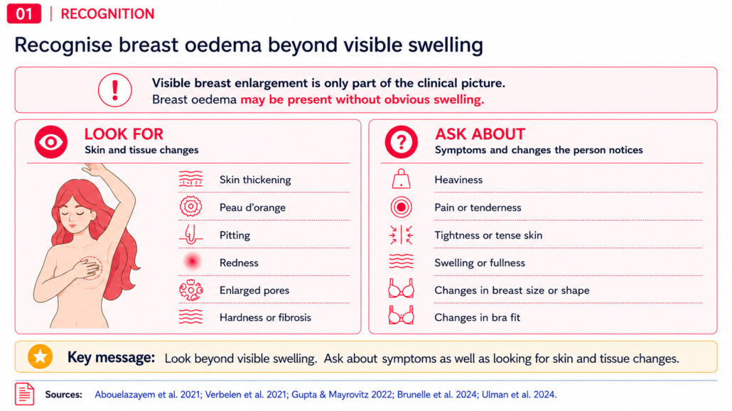
Figure 1. Recognising breast oedema beyond visible swelling. Patient-reported symptoms and skin or tissue changes can provide complementary information when visible enlargement is absent.
What has this research added?
Recognition of these wider signs and symptoms may help clinicians identify breast oedema that could otherwise be overlooked. Asking about heaviness, pain, tissue or skin changes, altered breast shape or size and changes in bra fit can provide information that visual examination alone may not capture.
How common is it?
Research published from 2019–2026 has shown that there is no single prevalence figure for breast oedema, but it has also helped explain why estimates differ and what those differences may mean clinically.
Importantly, reported prevalence is strongly influenced by how breast oedema is defined, how it is assessed and when assessment occurs. In the 2026 systematic review by Barletti et al., clinically assessed prevalence ranged from 2.7% to 24.8%, while some ultrasound-based studies identified breast tissue changes in substantially greater proportions of women. The review noted that objective measures such as ultrasound and TDC can identify tissue changes that may not be apparent on physical examination alone.[6,15]
A lower reported prevalence may reflect differences in case definition, assessment method or timing rather than a genuinely lower frequency of breast oedema. Young-Afat et al. similarly noted that differences between prevalence studies could be explained in part by different patient-reported measures, definitions and study designs.[7]
When assessment occurs is equally important. Breast oedema can already be present before radiotherapy, may become more evident during the months following treatment and may subsequently improve in many women.[7,24] Longitudinal studies therefore show that prevalence is not static but changes across the treatment and recovery period.
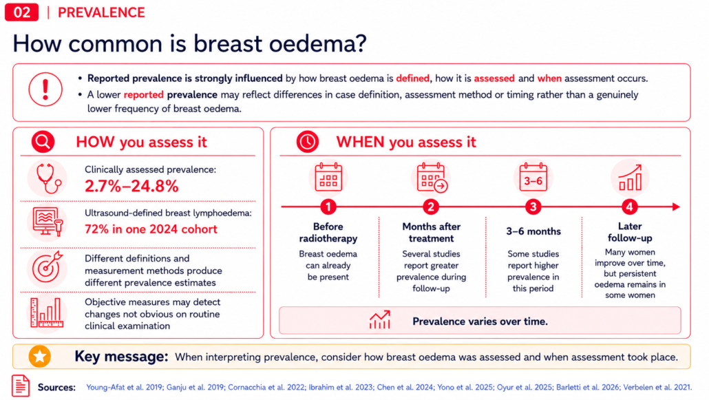
Figure 2. Interpreting reported prevalence of breast oedema. Estimates vary according to case definition, assessment method and timing after treatment.
The practical message is not to select one prevalence figure as the answer to “How common is breast oedema?” Instead, consider what was assessed, how it was assessed and when the assessment took place.
Risk factors and change over time
Across studies, characteristics associated with breast oedema have included more extensive axillary treatment, larger breast size or volume, higher BMI, some radiotherapy characteristics, chemotherapy in some cohorts, postoperative cellulitis and concurrent arm lymphoedema.[7,9–12,15]
These associations do not allow reliable prediction for an individual woman, but they do help identify circumstances in which greater awareness and follow-up may be useful.
Research has also improved our understanding of when breast oedema may occur and how it may change over time. Breast oedema can already be present before radiotherapy, several studies report greater prevalence during the months following treatment, and symptoms improve spontaneously over subsequent months in many women.[7,12,24,26]
Recent radiotherapy-specific studies suggest the relationship is more nuanced than fractionation alone. Breast or treatment volume, nodal irradiation and boost-related factors are being investigated as potential contributors, while one large 2025 cohort found no significant difference in breast oedema between normofractionated and hypofractionated schedules.[12,14]
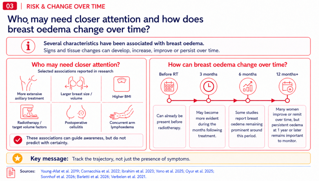
Figure 3. Factors associated with breast oedema and change over time. Associations may guide clinical awareness; time points are illustrative and individual trajectories vary.
What has this research added?
Breast oedema is increasingly understood as a dynamic condition rather than a single post-treatment event.
Monitoring whether symptoms are improving, persisting or worsening over time may provide more useful clinical context than simply recording whether symptoms are present.[24]
Improving assessment
Another important area of progress has been the development and investigation of more structured ways to assess breast oedema.
The Breast Edema Questionnaire (BrEQ) was developed to capture breast oedema symptoms and their effect on daily functioning; the initial clinimetric validation focused on the Part 1 symptom score.[5] Objective approaches including ultrasound measurement of dermal thickness and TDC have also been investigated, while ICG lymphography has provided new insight into altered lymphatic drainage patterns following breast cancer treatment.[1–5]
Fearn et al.’s systematic review found that ultrasound measurement of dermal thickness had the strongest measurement-property evidence among the clinician-reported approaches evaluated. TDC and several patient-reported measures were promising but required further validation.[2] Subsequent work by Riches et al. found that both ultrasound and TDC could distinguish between oedematous and non-oedematous breast tissue.[4]
More recent work has also begun to explore multiparameter classification, combining ultrasound findings with physical examination and patient-reported outcomes rather than relying on one measure alone.[6]
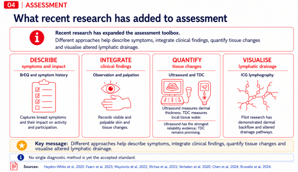
Figure 4. Recent developments in breast-oedema assessment. Symptoms, clinical findings, quantitative measures and specialist lymphatic imaging provide different forms of information; no single diagnostic standard has yet been established.
What has this research added?
Recent research has strengthened the assessment of breast oedema by providing more structured ways to capture symptoms and their impact,[5] integrate clinical findings with other assessment information,[6,26] quantify tissue changes using ultrasound and TDC,[2–4,6] and visualise altered lymphatic drainage with ICG lymphography.[1]
Together, these approaches can provide complementary information when building an assessment picture, rather than relying on any one source of information alone.[1,2,4,6,26]
No single diagnostic method is yet the accepted standard, but the research gives clinicians several complementary ways to describe, document and measure different aspects of breast oedema.[2,26]
Understanding the impact on women
Research has also moved beyond measuring breast oedema to examining what it means for the women experiencing it.
Young-Afat et al. found that breast oedema was independently associated with greater breast pain and with poorer quality of life, physical functioning and body image at some assessment points.[7] Their findings helped establish that the impact of breast oedema extends beyond measurable breast changes.
Ulman et al. added women’s own experiences. Participants described unfamiliar and distressing symptoms, effects on movement, clothing and bra choice, emotional and social consequences, and uncertainty about what was happening. Many felt unprepared for breast or trunk lymphoedema, and some experienced delays in diagnosis or treatment when their concerns were not recognised.[16]
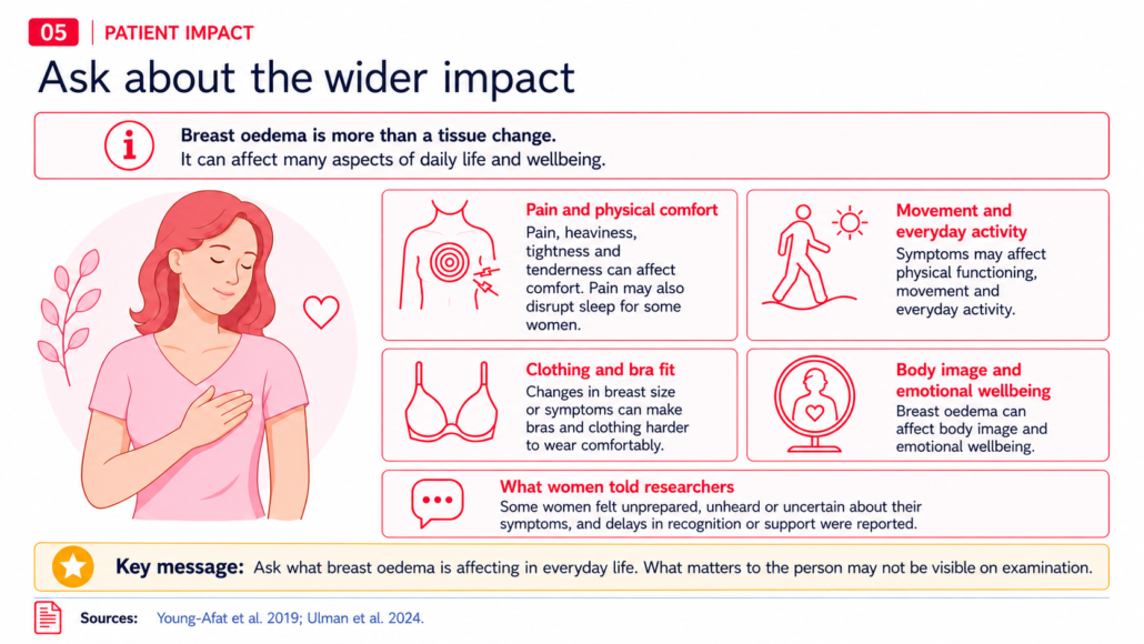
Figure 5. Wider patient impact of breast oedema. Symptoms can affect comfort, physical function, clothing, body image, emotional wellbeing and daily life.
What has this research added?
Recent research has made the wider impact of breast oedema much more visible. It can affect pain, physical functioning, clothing and bra fit, body image, emotional wellbeing and everyday life.[7,16]
It also shows why the woman’s experience matters clinically. Asking what has changed, what symptoms are affecting and what concerns the person has may reveal impacts that are not apparent from examination alone.[7,16]
What have we learned about management?
Over the 2019–2026 period, breast-specific intervention research has also begun to provide information that was previously limited.
Kilbreath et al.’s randomised trial found that a 12-week combined aerobic and resistance exercise program was safe and was associated with greater improvement in breast-related symptoms than usual care, without exacerbating lymphoedema.[19]
Compression research has produced more mixed findings.[17,18,21] Gregorowitsch et al.’s pilot study of a compression vest reported improvements in breast-oedema symptoms, although its uncontrolled design limits conclusions about treatment efficacy.[17] Johansson et al. found no significant advantage of a compression sports bra over a standard bra, although both groups improved over time.[18] More recently, Williams and Muldoon’s pilot audit reported improvements in symptoms and comfort with the Solidea Silver Wave Skin Bra, although its uncontrolled design means treatment efficacy cannot be established from the audit alone.[21]
Research into manual lymphatic drainage is also progressing. Haspolat et al. examined acute changes following MLD and compression with exercise,[20] while the preliminary 2026 randomised trial by Erkol et al. reported greater improvements in several outcomes when MLD was added to education, compression and exercise. The authors emphasised that larger studies are needed.[22]
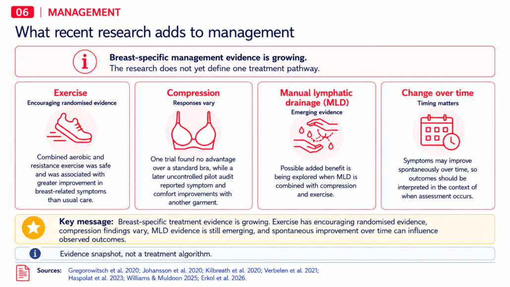
Figure 6. Current breast-specific management evidence. Exercise has encouraging randomised evidence, findings for compression vary and evidence for MLD remains preliminary; spontaneous improvement should be considered when interpreting treatment response.
What has this research added?
The intervention studies do not yet establish one treatment pathway, but they have begun to provide breast-specific evidence rather than relying solely on approaches extrapolated from limb lymphoedema.[17–22]
Exercise has encouraging randomised evidence, findings for compression remain mixed, and evidence for MLD remains preliminary.[17–22]
Spontaneous improvement over time also needs to be considered when treatment outcomes are interpreted.[18,24] Symptoms may decline during the months following radiotherapy as part of the natural course of breast oedema, making it important to distinguish treatment effect from improvement that may have occurred over time.
What does research from 2019–2026 add to clinical practice?
The studies reviewed do not provide a single diagnostic or treatment protocol, but together they offer several practical insights.
- Look beyond visible swelling. Breast oedema may also present with pain, heaviness, skin change, hardness and altered tissue texture.[23–26]
- Ask about the person’s experience. Research demonstrates effects on physical function, body image, quality of life and everyday activities, as well as the distress that can occur when symptoms are not recognised.[7,16]
- Consider timing and trajectory. Breast oedema may change considerably during the months following treatment, including spontaneous improvement in some women.[7,12,24]
- Draw on complementary information when assessing. Patient-reported symptoms, clinical examination and objective measures each contribute different information.[1,4–6,26] Among the measurement approaches reviewed, ultrasound dermal thickness currently has the strongest reliability evidence.[2]
- Interpret treatment response in the context of the evidence. Exercise has encouraging randomised evidence, compression findings are mixed and MLD evidence is continuing to develop.[17–22]
The picture in 2026
Research published since 2019 has changed what we know about breast oedema after breast cancer treatment.
Breast oedema is now better characterised as a condition that may involve pain, heaviness, skin and tissue changes as well as swelling. Research has provided greater insight into when it occurs and how it changes over time, identified recurring factors associated with its development, and demonstrated its impact on physical function, quality of life and body image.
Assessment has also progressed. A breast-specific patient-reported measure has been developed, ultrasound has demonstrated useful reliability, TDC continues to show promise and ICG lymphography has provided new information about altered breast lymphatic drainage.
The treatment evidence base remains smaller, but it too is developing. Randomised evidence supports the safety of aerobic and resistance exercise, compression studies have helped clarify the limitations and possibilities of current approaches, and recent research is beginning to explore the potential contribution of MLD.
While most prevalence and risk-factor evidence remains observational, and treatment trials remain relatively small, research published from 2019–2026 has given clinicians a much stronger foundation for recognising breast oedema, asking the right questions, monitoring change over time and making evidence-informed management decisions.
Further research is still needed to establish consistent diagnostic criteria and determine which treatments provide the greatest benefit for which women, but the clinical picture in 2026 is considerably clearer than it was in 2019.
References and further reading
Most of the references below include a link to the full article where available. Click the article title to open it in a new window.
| Ref. | Article | Open access? | DOI / abstract link |
Assessment and diagnosis |
|||
| 1 | Heydon-White et al. (2020), Assessing breast lymphoedema following breast cancer treatment using indocyanine green lymphography | No | 10.1007/s10549-020-05661-y |
| 2 | Fearn et al. (2023), Quantification of breast lymphoedema following conservative breast cancer treatment: a systematic review | Yes | 10.1007/s11764-022-01278-w |
| 3 | Mayrovitz et al. (2022), Breast tissue dielectric constant as a potential breast edema assessment parameter | No | 10.1089/lrb.2020.0137 |
| 4 | Riches et al. (2023), Improving the assessment and diagnosis of breast lymphedema after treatment for breast cancer | Yes | 10.3390/cancers15061758 |
| 5 | Verbelen et al. (2020), Development and clinimetric properties of the Dutch Breast Edema Questionnaire (BrEQ-Dutch version) | No | 10.1007/s11136-019-02337-z |
| 6 | Chen et al. (2024), Classification of breast lymphedema in a racially diverse cohort | No | PubMed abstract |
Prevalence, risk factors and change over time |
|||
| 7 | Young-Afat et al. (2019), Breast edema following breast-conserving surgery and radiotherapy: patient-reported prevalence, determinants, and effect on health-related quality of life | Yes | 10.1093/jncics/pkz011 |
| 8 | Ganju et al. (2019), Incidence of breast lymphedema and predictors of its development in patients receiving whole breast radiation therapy after breast-conservation surgery | No | PubMed abstract |
| 9 | Cornacchia et al. (2022), Breast edema after conservative surgery for early-stage breast cancer: retrospective single-center assessment of risk factors | No | PubMed abstract |
| 10 | Ibrahim et al. (2023), Iatrogenic breast lymphoedema: incidence, diagnosis, and associated risk factors | No | 10.1007/s13193-023-01725-9 |
| 11 | Yono et al. (2025), Factors associated with breast lymphedema after adjuvant radiation therapy in women undergoing breast conservation therapy | Yes | 10.1016/j.breast.2024.103846 |
| 12 | Oyur et al. (2025), Breast edema after breast-conserving surgery and radiotherapy: introduction of a clinically meaningful classification and evaluation of the incidence after normo- and hypofractionated treatments | Yes | 10.3390/cancers17142368 |
| 13 | Shaw et al. (2026), Real world incidence of breast lymphoedema following breast conserving surgery and different radiotherapy schedules – conference abstract | No | 10.1016/j.ejso.2025.111210 |
| 14 | Sonnhoff et al. (2026), Association between radiation volume and breast density for skin toxicity and breast edema after radiotherapy in breast conserving therapy of breast cancer | Yes | 10.1186/s13014-026-02833-w |
| 15 | Barletti et al. (2026), Breast edema in women with arm lymphedema following breast-conserving surgery: a systematic review | Yes | 10.7759/cureus.108129 |
Patient experience and clinical impact |
|||
| 16 | Ulman et al. (2024), Exploring women’s experiences of breast or trunk lymphoedema following treatment for breast cancer | Yes | 10.1080/07347332.2023.2218374 |
Management |
|||
| 17 | Gregorowitsch et al. (2020), Compression vest treatment for symptomatic breast edema in women treated for breast cancer: a pilot study | No | 10.1089/lrb.2018.0067 |
| 18 | Johansson et al. (2020), Compression treatment of breast edema: a randomized controlled pilot study | No | 10.1089/lrb.2018.0064 |
| 19 | Kilbreath et al. (2020), Reduction of breast lymphoedema secondary to breast cancer: a randomised controlled exercise trial | No | 10.1007/s10549-020-05863-4 |
| 20 | Haspolat et al. (2023), Acute effects of manual lymphatic drainage and compression with exercise therapy on breast lymphedema following breast-conserving surgery and radiotherapy | No | 10.1089/lrb.2022.0037 |
| 21 | Williams & Muldoon (2025), A pilot audit of the Solidea® Silver Wave Skin Bra for women with breast and trunk lymphoedema after breast cancer treatment | No | Wounds UK article & abstract |
| 22 | Erkol et al. (2026), Effect of manual lymph drainage on breast edema after breast-conserving surgery and radiotherapy: a preliminary randomized controlled trial | Yes | 10.3390/cancers18101510 |
Reviews and broader perspectives |
|||
| 23 | Abouelazayem et al. (2021), Breast lymphedema after conservative breast surgery: an up-to-date systematic review | No | 10.1016/j.clbc.2020.11.017 |
| 24 | Verbelen et al. (2021), Breast edema, from diagnosis to treatment: state of the art | Yes | 10.1186/s40945-021-00103-4 |
| 25 | Gupta & Mayrovitz (2022), The breast edema enigma: features, diagnosis, treatment, and recommendations | Yes | 10.7759/cureus.23797 |
| 26 | Brunelle et al. (2024), Breast lymphedema following breast-conserving treatment for breast cancer: current status and future directions | No | 10.1007/s10549-023-07161-1 |
Transparency statement: This article was developed with the support of AI to assist in collating and synthesising the published research listed above. Research findings and conclusions have been attributed to their original sources, with the content reviewed and edited for accuracy and clinical relevance.
Acknowledging our Industry Partners
LES acknowledges and thanks Essity, Hartman and OPC Health for supporting the development of this resource and ongoing clinical learning. Inclusion does not imply endorsement, and clinicians are encouraged to apply their own professional judgement in all aspects of patient care.